
⚠ Possible eye emergency
Sudden new flashes, a major increase in floaters, or a curtain/shadow in vision may require urgent eye evaluation. Do not wait for a routine appointment — call the clinic on +91 93729 47075 or seek urgent eye care.
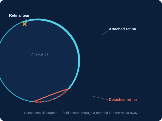
What Is a Retinal Detachment?
The retina lies against the inside back wall of the eye like wallpaper. A retinal detachment occurs when the retina lifts away from that wall, separating it from the layer that supplies it with oxygen and nutrients. Cut off from its blood supply, the detached retina stops working, and its cells begin to die.
The commonest type (rhegmatogenous detachment) begins with a retinal tear: the vitreous gel inside the eye shrinks with age, tugs on the retina and tears it. Fluid then passes through the tear and lifts the retina off. Less commonly, scar tissue pulls the retina off (tractional detachment, seen in advanced diabetic disease) or fluid collects under it without a tear (exudative detachment).
Warning Symptoms
Retinal detachment is painless, which makes its warning signs easy to dismiss. Take them seriously: a sudden shower of new floaters — many dark spots, threads or cobwebs appearing over hours or days; repeated flashes of light (photopsia), especially in the side vision; and a dark curtain, veil or shadow spreading across any part of the visual field. Blurring of central vision means the macula is threatened or already involved.
These symptoms can also occur with a retinal tear before detachment — the stage at which a simple laser procedure can often prevent the detachment altogether.
Who Is at Risk?
Risk is higher in people with moderate to high myopia (minus power), those who have had cataract or other eye surgery, anyone with a previous detachment or tear in either eye, people with a family history of retinal detachment, those who have had significant eye trauma, and patients with advanced diabetic retinopathy. Ageing itself increases risk as the vitreous naturally shrinks.
How It Is Diagnosed
Diagnosis is made with a dilated retinal examination, often with indirect ophthalmoscopy that lets the surgeon view the far periphery of the retina. If bleeding inside the eye blocks the view, an ultrasound (B-scan) shows whether the retina is attached. The examination also maps the location and number of retinal tears, which guides the surgical plan.
Treatment Concepts
A retinal tear without detachment is usually sealed with laser photocoagulation around the tear — a short outpatient procedure. An established detachment needs surgery: options include pneumatic retinopexy (a gas bubble plus laser or freezing treatment), scleral buckling (a silicone band supporting the eye wall from outside), or vitrectomy (removing the vitreous gel, flattening the retina and holding it with gas or silicone oil). The choice depends on the tear pattern, lens status and surgeon's assessment.
Surgery reattaches the retina in the large majority of cases, sometimes requiring more than one procedure. Visual recovery depends heavily on whether the macula was still attached at the time of surgery — which is why hours and days matter.
Recovery Expectations
After surgery, patients may need to keep a specific head position for some days so the gas bubble supports the repaired retina. Vision typically improves gradually over weeks to months; it rarely returns instantly. Air travel is forbidden while gas remains in the eye. Follow-up visits monitor healing, eye pressure and the other eye, which carries its own lifetime risk and is examined carefully.
Understanding the diagnosis
A diagnosis of Retinal Detachment can raise practical questions about symptoms, tests, treatment and recovery. A careful consultation begins with your story: what you have noticed, how quickly it appeared, whether one or both eyes are involved, and whether conditions such as diabetes, blood pressure, previous surgery or injury may affect the eye. The aim is not to rush to a procedure. It is to understand the health of the whole eye and explain which findings matter most for your vision.
Symptoms and when to seek help
Some eye conditions develop slowly and may be noticed only during a routine examination. Others can cause a sudden change in sight. New flashes, a sudden shower of floaters, a curtain or shadow, sudden loss of vision, severe pain, marked redness after injury or rapidly worsening symptoms should be treated as urgent concerns. Routine appointments are appropriate for stable symptoms, but waiting is not a substitute for evaluation when vision changes quickly.
How the consultation works
Your visit is structured around a detailed history and an examination of each eye. Depending on the concern, this can include refraction, eye pressure measurement, slit-lamp examination, dilated retinal examination, retinal photography, OCT, visual fields, biometry, corneal mapping or other tests. Not every patient needs every test. The reason for each test should be clear, and you should have time to ask what the result means for your own situation.
Why measurements matter
Treatment decisions in retina and vitreous care depend on measurements as well as symptoms. Two people with similar glasses powers or similar complaints may have very different corneal, lens, retinal or optic-nerve findings. Good planning therefore combines examination findings, imaging, medical history, work demands, family history and personal priorities. Measurements are useful when they lead to a safer, more individualised decision rather than being treated as a sales promise.
Treatment options are individual
There may be more than one reasonable way to manage Retinal Detachment. Options can include observation with scheduled monitoring, medicines, laser treatment, injections, glasses, contact lenses or surgery. The right option depends on the stage of disease, the health of the other eye, your general health, likely benefits, possible risks, cost, recovery requirements and how urgently treatment is needed. A consultation should explain alternatives, not only the most intensive intervention.
Preparing for an appointment
Bring previous eye reports, scans, prescriptions, medication lists and details of any diabetes, hypertension or other long-term condition. If you use contact lenses, ask whether they should be removed before measurements. Dilating drops can temporarily blur near vision and make bright light uncomfortable, so arrange transport if your clinician advises dilation. Write down your main questions. Clear questions help you and the clinical team use the appointment well.
What recovery may involve
Recovery varies by diagnosis and treatment. Some visits involve no downtime, while others require drops, activity restrictions, protective eyewear or several follow-up examinations. It is important to use medicines exactly as prescribed, avoid rubbing the eye, attend reviews and contact the clinic if pain, redness, discharge or vision changes are unexpected. Do not compare your recovery with someone else’s; the treatment and the eye are personal.
Long-term monitoring
Many eye conditions are managed over time rather than solved in one visit. Follow-up allows the doctor to compare vision, pressure, scans and symptoms with earlier findings. Keep a record of appointments and medicines, and tell the clinic about changes in general health. People living with diabetes or blood-pressure problems should continue their regular medical care because systemic health and eye health are closely connected.
Questions to ask your doctor
Useful questions include: What exactly did the examination show? What is the goal of treatment? What happens if we monitor for now? Which alternatives apply to my eye? What are the common and serious risks? How many follow-ups will I need? Which symptoms should prompt an urgent call? What should I avoid during recovery? A good decision is one you understand and can follow safely.
A patient-first approach
At Sentra Clinic & Hospital, the emphasis is on a calm explanation, appropriate investigations and a plan that fits the patient. The website cannot diagnose an eye condition, confirm eligibility for surgery or predict an individual result. Those decisions require an in-person examination. Educational information can help you prepare, but it should support—not replace—the conversation with a qualified eye specialist.
Access and follow-up in Malad East
Dr. Rohit Modi consults at Sentra Clinic & Hospital at Shah Arcade 2, 002, First Floor, B Wing, Rani Sati Road, Malad East, Mumbai 400097. Patients can call 9372947075 or 02246057489 for appointments and clinic guidance. If you are travelling from another part of Mumbai, confirm the appointment time before leaving and bring your reports so the consultation can stay focused on your needs.
Medical disclaimer
This page is for patient education and general awareness. It does not create a doctor–patient relationship and does not replace diagnosis, treatment or emergency care. Results, risks and recovery differ between individuals. If you have a sudden or severe change in vision, seek urgent professional eye care rather than waiting for an online response or a routine appointment.
Understanding the diagnosis
A diagnosis of Retinal Detachment can raise practical questions about symptoms, tests, treatment and recovery. A careful consultation begins with your story: what you have noticed, how quickly it appeared, whether one or both eyes are involved, and whether conditions such as diabetes, blood pressure, previous surgery or injury may affect the eye. The aim is not to rush to a procedure. It is to understand the health of the whole eye and explain which findings matter most for your vision.
Symptoms and when to seek help
Some eye conditions develop slowly and may be noticed only during a routine examination. Others can cause a sudden change in sight. New flashes, a sudden shower of floaters, a curtain or shadow, sudden loss of vision, severe pain, marked redness after injury or rapidly worsening symptoms should be treated as urgent concerns. Routine appointments are appropriate for stable symptoms, but waiting is not a substitute for evaluation when vision changes quickly.
How the consultation works
Your visit is structured around a detailed history and an examination of each eye. Depending on the concern, this can include refraction, eye pressure measurement, slit-lamp examination, dilated retinal examination, retinal photography, OCT, visual fields, biometry, corneal mapping or other tests. Not every patient needs every test. The reason for each test should be clear, and you should have time to ask what the result means for your own situation.
Why measurements matter
Treatment decisions in retina and vitreous care depend on measurements as well as symptoms. Two people with similar glasses powers or similar complaints may have very different corneal, lens, retinal or optic-nerve findings. Good planning therefore combines examination findings, imaging, medical history, work demands, family history and personal priorities. Measurements are useful when they lead to a safer, more individualised decision rather than being treated as a sales promise.
Treatment options are individual
There may be more than one reasonable way to manage Retinal Detachment. Options can include observation with scheduled monitoring, medicines, laser treatment, injections, glasses, contact lenses or surgery. The right option depends on the stage of disease, the health of the other eye, your general health, likely benefits, possible risks, cost, recovery requirements and how urgently treatment is needed. A consultation should explain alternatives, not only the most intensive intervention.
Preparing for an appointment
Bring previous eye reports, scans, prescriptions, medication lists and details of any diabetes, hypertension or other long-term condition. If you use contact lenses, ask whether they should be removed before measurements. Dilating drops can temporarily blur near vision and make bright light uncomfortable, so arrange transport if your clinician advises dilation. Write down your main questions. Clear questions help you and the clinical team use the appointment well.
What recovery may involve
Recovery varies by diagnosis and treatment. Some visits involve no downtime, while others require drops, activity restrictions, protective eyewear or several follow-up examinations. It is important to use medicines exactly as prescribed, avoid rubbing the eye, attend reviews and contact the clinic if pain, redness, discharge or vision changes are unexpected. Do not compare your recovery with someone else’s; the treatment and the eye are personal.
Long-term monitoring
Many eye conditions are managed over time rather than solved in one visit. Follow-up allows the doctor to compare vision, pressure, scans and symptoms with earlier findings. Keep a record of appointments and medicines, and tell the clinic about changes in general health. People living with diabetes or blood-pressure problems should continue their regular medical care because systemic health and eye health are closely connected.
Questions to ask your doctor
Useful questions include: What exactly did the examination show? What is the goal of treatment? What happens if we monitor for now? Which alternatives apply to my eye? What are the common and serious risks? How many follow-ups will I need? Which symptoms should prompt an urgent call? What should I avoid during recovery? A good decision is one you understand and can follow safely.
A patient-first approach
At Sentra Clinic & Hospital, the emphasis is on a calm explanation, appropriate investigations and a plan that fits the patient. The website cannot diagnose an eye condition, confirm eligibility for surgery or predict an individual result. Those decisions require an in-person examination. Educational information can help you prepare, but it should support—not replace—the conversation with a qualified eye specialist.

Eye Surgeon | Refractive & Retina Specialist · FRCS (Glasgow)
Last reviewed: 4 September 2026